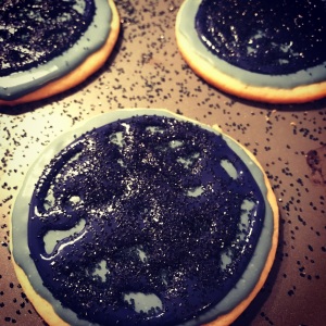
Okay, that was disturbing. But it was a lot of fun making cookies in the shape of different blood cells for our lectures on anemia and leukemia this week!
The colors didn’t always turn out very appetizing – I was going for realness, though, and I think I nailed it! Shall we review our different hematopathologic diseases? I think we shall.

Sickle cell anemia
Here’s sickle cell anemia, with sickle cells (obviously), and a post-splenectomy blood picture including target cells, spherocytes, Howell-Jolly bodies, nucleated red cells, and a thrombocytosis. Basically, everything that the spleen normally filters from the blood – nucleated red cells, Howell-Jolly bodies, Pappenheimer bodies (which are little iron granules), target cells, and spherocytes – gets out into the blood when you don’t have a spleen. The spleen also holds about 1/3 of your platelets at any given time – so removing the spleen removes their little home, and they have to wander the streets (blood vessels), begging for food. So sad.

Microangiopathic hemolytic anemia
Here we have helmet cells, spherocytes, and the most specific (but unimaginatively-named) schistocyte of all: the triangulocyte.

Megaloblastic anemia
In this one, you’ve got oval macrocytes (gigantic, oval-shaped red cells) and hypersegmented neutrophils.

Chronic myeloid leukemia
Here we have a neutrophilic leukocytosis with a left shift (there’s a myelocyte, top left, a metamyelocyte, bottom left, and a promyelocyte, center) and a basophilia (right). Check out how mature the chromatin is in the myelocyte, metamyelocyte, and segmented neutrophil – but how fine it is (you can even see nucleoli!) in the promyelocyte). Also, the granules in the promyelocyte are in the cytoplasm and over the nucleus as well. The basophil granules are kinda obscuring the nucleus, but that’s what happens in real life, so we’ll call it artistic rendering.

Myeloblast with Auer rod
Not so appetizing, color-wise, but pretty accurate, microscope-wise. Check out the Auer rod next to the nucleus (which, by the way has such fine chromatin that you can see nucleoli through it). The Auer rod is a linear aggregation of primary (azurophilic) granules that only happens in malignant myeloblasts.
 Faggot cell
No, we’re not using the term that way – we’re talking about the old English for “bundle of sticks” (which is where the British slang for a cigarette, “fag,” came from). There are a TON of Auer rods in faggot cells – way more than I could draw here.
Chronic lymphocytic leukemia
In chronic lymphocytic leukemia, the lymphocytes are small, with clumped chromatin and not much else going on. They look like regular old mature lymphocytes. So I thought they deserved a light dusting of sprinkles. Not particularly appropriate histologically, but cute.
 In the classroom
Here’s some of the cookies in the classroom (the Reed Sternberg cells were a big hit, so that tray’s already empty).
Final touches
And here’s a shot of part of the craziness at the frosting stage. It was one of those projects that kind of scaled up in size very quickly! This is about 1/3 of all of the cookies…


You are the most awesome teacher I have ever heard of! keep on teaching 😀
Awww thanks Mohammed!! I will definitely keep on teaching 🙂
Hello Kristine,
What an awesome idea… I might ‘steal’ it for next year when teaching the med students about general pathology. I keep referring to foodstuff when lecturing/tutoring. This will add another dimension to it. I can just imagine their faces when we talk about liquefactive necrosis and vanilla custard!!!
Cheers
Dwight
UTas
Hi Dwight! Wow – yeah, that would be a great dessert! Pathology and food go so well together 🙂
Kristine, awesome , most of the teachers teach us by looking into the book pictures, while you are awesome in every way….
keep it up !
Its really creative ma’am!!! You are Always best !!!!
This is beyond amazing. I wish you were my professor! (But I suppose I’ll settle for your phenomenal website and emails 🙂
This is awesome. Wish we had more creative teachers like you!
Thanks, Lizzy!! If you email me your address, I’ll send you some next time I make them! 🙂
What a delicious way to make students remember hematology. i wish i would be your student.
if you allow me i may share these cookies with my students. but i am afraid they will ask me to make real ones for them…
It’s very easy 🙂 I’ll email you the recipe if you want!
Thank you Kristine. Sure. i will wait. with best regards
I got here after a few hours spent browsing the web for resources on chilblain lupus (being one of 70 patients on the planet ever diagnosed with this crap can be pretty frustrating :-/ ) and while looking for articles about immunofluorescence, I stumbled upon your website. Wow, you are amazing! JUST AWESOME!!! I wish I was young again and could become your student ;))
Awesome idea
Kristine: I agree with all of the other comments her that you are a phenomenal teacher and very creative as well! What a great idea and a very visual way for your students to remember these cells forever.
Lots of people are brilliant but not many can take that brilliance and share it with others (teach) like you do! Just because you have a PhD or an MD does not mean you are a good teacher.
I too wish I had you as a teacher, but I’m happy for your students! You make me wish I had studied Pathology more than Hene/Onc but I had to learn a ton of Path too to be a great physician.
You have a nice way of taking complex topics and making them easily understandable and able to therefore memorize! All physicians must be teachers, if not to Med students, at least to all of our Pts!
Thanks again for all you do to enhance understanding of Pathology.
Your blood cookies are simply amazing!
God Bless you!
Dr. Krafts:
Is there any way to receive your recipes for the Blood Cookies? I appreciate it. Thank you and God Bless you!
Sure! I use this recipe for my cookies, and this recipe for the icing.