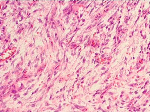
A 25 year-old male presents with a mass on the volar aspect of his forearm. He first noticed the lesion 2 weeks ago, when he developed pain in his arm, and since then it has grown rapidly to reach its current size of approximately 5 cm.
What is the diagnosis?
A. Lipoma
B. Nodular fasciitis
C. Neurofibroma
D. Myxoid liposarcoma
E. Osteosarcoma
(Scroll down for the answer)
Ahh this diagnosis! Many a first-year pathology resident has missed this diagnosis during “unknown conference” (a particularly painful exercise in which the attendings put out a tray of unknown slides one evening, and then the next day, you have a conference where you go around the room and everyone gives their diagnosis for each slide). It’s kind of embarrassing missing a diagnosis – but the good thing is that the next time you see it, you’ll remember it for sure. Lucky for you, we’re doing this online, not in a dark room in front of all of your colleagues. So if you miss it this time, don’t worry – the next time you see it, you’ll nail it!
The diagnosis in this case is nodular fasciitis. Nodular fasciitis is a benign disorder which most commonly occurs in adults, and most commonly is found on the volar aspect of the forearm. It’s thought to be an over-reaction to trauma (though if you ask patients, many will not remember any preceding trauma).
So why is it such a tricky diagnosis for young residents? Because it looks horrible! You’d swear it was some sort of myxoid sarcoma thing – but it’s totally benign. It is composed of plump spindle-shaped fibroblasts with conspicuous nuclei arranged in a myxoid background, usually with some small, thin-walled blood vessels, extravasated red cells, and scattered lymphocytes. Which in and of itself is scary-looking. But in addition, it’s hypercellular, and it has increased mitotic activity – two things you see often in sarcoma.
One point I’d like to make about mitoses: just seeing mitoses is not, in and of itself, diagnostic of malignancy. Lots of benign tumors have an increased mitotic rate. Malignant tumors tend to have more mitoses than benign tumors – but this doesn’t always hold true. Even normal tissues (like GI epithelium) can have mitotic figures. So while sometimes helpful in the diagnosis of tumors, in reality, the presence of mitoses just means that the thing, whatever it is, is making new cells.
If you see an atypical mitosis, however, then you worry. One of the most significant and characteristic forms of atypical mitosis is the tri-polar (or pentapolar, or septapolar, though those are less common) mitosis. Normal mitoses do not have an odd number of poles! If you see one of those babies, it’s a malignancy until proven otherwise (and it probably won’t be).
Back to the lesion. The prognosis is excellent for patients with this diagnosis. Excision is curative.
If you want to test yourself with other unknown cases, here are some to try:
- Case 1: 20-year-old male who died suddenly
- Case 2: 72-year-old male with right calf mass
- Case 3: 67-year-old female with pancytopenia
- Case 4: 59-year-old male with severe headaches
- Case 5: 38-year-old female with deep venous thrombi
- Case 6: 13-year-old male with cerebellar mass
- Case 7: 45-year-old male with pulmonary emphysema
- Case 8: 38-year-old male with AIDS and headaches


Thank you. I got the idea.
I am pathologist and reading your own biopsy taken from something that has grown so rapidly in 5 days time from the volar aspect of my right forearm 9 days ago was a nightmare…the 2 cm mass was removed piece meal…I showed the slide to our junior consultant in our department and he was worried about the “infiltrating” behavior of the “tumor” into the subcutaneous fat..and he suggested it be referred to a near emeritus age pathologist staff who diagnosed it like this..”benign vascular tumor compatible with hemangioepithelioma”…it was a relief that it was benign..but I still believe that I am correct with my diagnosis, i.e. nodular fasciitis..the wound so far is undergoing healing and praying it never comes back again..and yes, I don’t recall ever having a trauma in that area..wishing I could post the picture of my forearm before and after the excision.
So sorry you had to go through that – sounds agonizing. Thanks for sharing your story!
update of my case: although the nodule returned a bit four days post excision(it was removed piece meal), it somehow is getting smaller..i have the belief that this reactive tumor do regress in time…but earlier my surgeon planned a fascial excision if it ever recurs..which means it will be a regional block anesthesia..omg!
oh btw, I haven’t told you that I have a giant cavernous hemangioma in my liver only found 3 years back…I underwent embolization for a planned resection but later, my surgeon retracted and studied the scans one more time..now she is suggesting transplant! well its been three years…at first I thought I can live with mild to moderate pain and this mechanically induced acid reflux due to tumor compression on my stomach…now I am thinking otherwise…but at the end of the day, I could not imagine life after transplant with all the immunosuppression and the possibility of rejection later…I am still hopeful that elsewhere, someone can study back my liver scans for I am amenable to resection alone…thankfully, I am a doctor and a pathologist and knows somehow what is happening inside me…oh there is so much id like to talk about and I am a bit off topic…anyway all the best for you Dr. Kristine. thanks much.
Hi there ,
Can you please describe how a liposarcoma would appear in histology and the differences from a lipoma ? Is nuclear scalloping classic for liposarcoma ?
thankyou